I am a PhD candidate in the Department of Electrical Engineering at Caltech, advised by Prof. Changhuei Yang. My current research focuses on:
- Computational microscopy - empowering imaging techniques with algorithms
- Synergizing microscopy, computation and artificial intelligence to advance life science research
My aspiration is to become an engineer-scientist who advances scientific discovery through innovative engineering designs.
I am currently on the 2025-2026 job market.
News
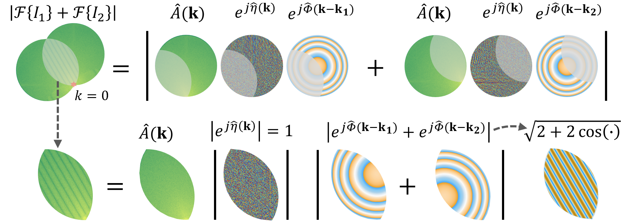
I presented our recent work on "Digital defocus aberration interference for autofocusing and digital refocusing in optical microscopy" at SPIE Photonics West 2026, San Francisco.
Our paper “Analytic Fourier ptychotomography for aberration-free and high-resolution volumetric refractive index imaging” is online at Nature Communications. Paper link.
I gave an invited talk on Fourier ptychographic microscopy at the International Webinar on Image & Graphics Technologies (Chinese Society for Image & Graphics, online).
I presented neural-field driven computational microscopy at the Optica Imaging Congress in Seattle.
Invited talk at Rice University (Houston) titled “Physics-based computational microscopy to advance life science research”.
ESE Seminar Series (Washington University in St. Louis): “Empowering microscopes with physics-based computation.” Talk page.
Computer Vision Seminar Series (University of Maryland, College Park): “Synergizing microscopy and computation to advance life science research.”
Our paper “Single-shot volumetric fluorescence imaging with neural fields” is online at Advanced Photonics. Paper link.
Awarded the Schmidt Academy for Software Engineering Graduate Research Fellowship supporting Fourier Ptychographic Microscopy software. Schmidt Academy GRA Fellows.
PNAS publishes “Investigating 3D microbial community dynamics of the rhizosphere using quantitative phase and fluorescence microscopy.” Paper · Science.org news.
“AI-guided histopathology predicts brain metastasis in lung cancer patients” is featured on the Caltech homepage. News.
“AI-guided histopathology predicts brain metastasis in lung cancer patients” is now online. Full list here.
I passed my candidacy exam.
Launched this personal homepage.
I joined the Caltech Biophotonics Lab.
Selected Publications
Full publication list here.


